


Multiple myeloma is not a single, uniform disease. It includes several types, distinguished by the proteins produced by cancerous plasma cells. Learning about the different types of multiple myeloma will help you better understand your diagnosis and the treatment options available to you.
What Is an Immunoglobulin?
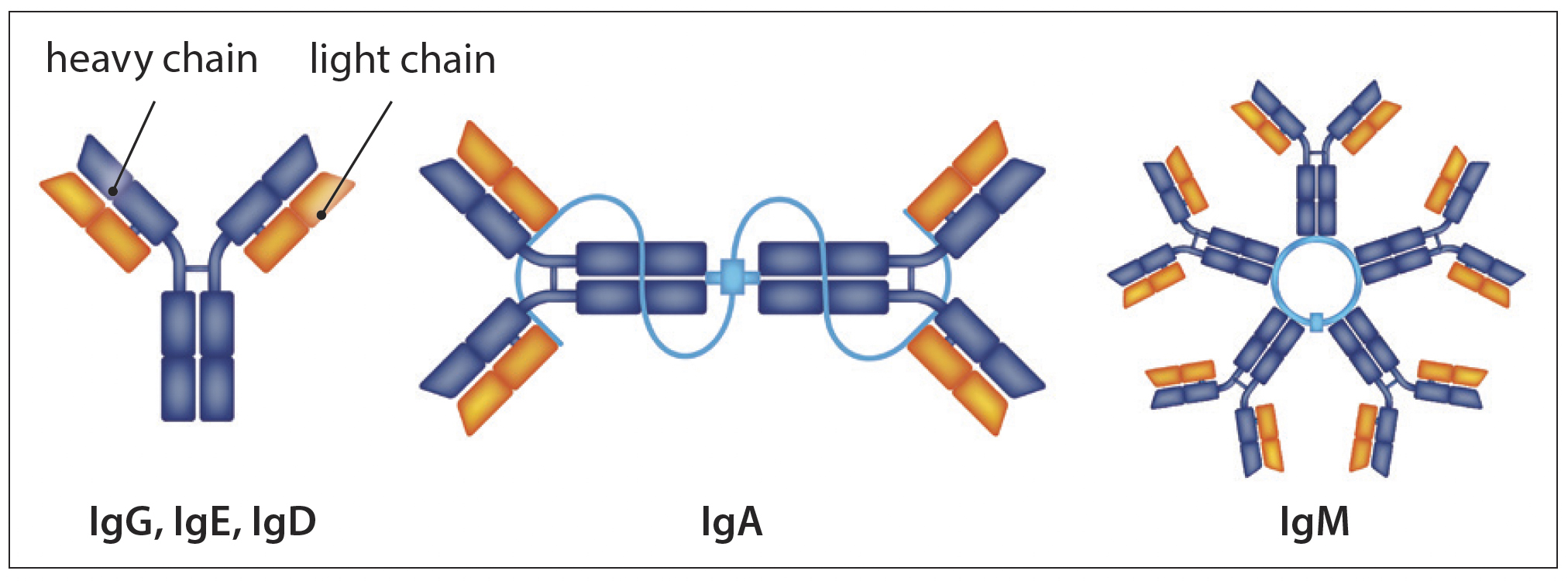
Each patient’s myeloma cells produce only 1 of the following 5 types of immunoglobulin proteins: IgG, IgA, IgD, IgE, and IgM. See the figure below.
Each immunoglobulin is made up of two heavy chains bound to two light chains. The two types of light chains are kappa (κ) and lambda (λ).
This figure shows the structure of immunoglobulins:
IgG myeloma
Approximately 65% of myeloma patients have IgG myeloma with either kappa or lambda light chains. The behavior of IgG myeloma conforms to CRAB criteria features.
IgA myeloma
IgA myeloma is the second-most common type, also with either kappa or lambda light chains. Patients with IgA myeloma sometimes have tumors outside of the bone.
IgD, IgE, or IgM myelomas
These three types of myeloma are quite rare. IgD myeloma can be accompanied by plasma cell leukemia (PCL).
Understanding Light Chains
Approximately 50% of myeloma patients produce free light chains in addition to the complete molecule combination of light chains bound to heavy chains.
About 20% of myeloma patients produce only light chains and no heavy chains. This is called Bence-Jones myeloma, named for the English doctor who first detected and identified light chains, and published his findings in 1848.
Light-chain M-proteins are smaller and weigh less than heavy chains, making it possible for them to fit through the tiny capillaries that send blood to the kidneys. The light chains that arrive by blood to the kidneys may build up to the point of blocking the kidney’s tubules, causing reduced kidney function.
This is why patients with light-chain only myeloma are more likely to have kidney damage or deposits of light chains or on nerves or other organs.
In the majority of patients with light-chain myeloma, their disease can be measured with the sFLC test.
In some patients, their myeloma can be measured only in the urine.
Only 1%–2% of myeloma patients produce very little or no M-protein of any type. These patients may have to be assessed by other means (e.g., imaging, bone marrow testing). This is often referred to as “non-secretory” myeloma.
The goal of treatment is to normalize the light-chain levels, especially the ratio, and to eliminate the M-protein.
Understanding the Types of Multiple Myeloma
Multiple myeloma can be classified in different ways, most commonly by the type of immunoglobulin the myeloma cells produce and the stage of the disease at diagnosis.
Classification by Immunoglobulin Type
Based on immunoglobulin type, there are different types of multiple myeloma, some more common than others:
- IgG myeloma: Approximately 65% of myeloma patients have IgG myeloma with either kappa or lambda light chains. The behavior of IgG myeloma conforms to CRAB criteria features.
- IgA myeloma: The second-most common type, with either kappa or lambda light chains. Patients with IgA myeloma sometimes have tumors outside of the bone.
- IgD, IgE, or IgM myelomas: These three types of myeloma are quite rare. If the type is IgM, it must be differentiated from Waldenstrom’s macroglobulinemia, a form of lymphoma that has an IgM monoclonal protein.
Classification by Disease Stage
Multiple myeloma may either be smoldering or active:
- Smoldering multiple myeloma (SMM): At this stage, the disease is present but quiet. You won't have any symptoms or damage to organs, such as the kidneys and bones. Recently, we have divided this into standard-risk and high-risk SMM, and some patients with high-risk SMM may be treated.
- Active multiple myeloma: When multiple myeloma is active, it presents symptoms such as bone pain and fatigue. It can also lead to organ damage, which may include bone lesions and kidney failure.
The specific immunoglobulin type of myeloma can manifest as either active or smoldering. For example, you may have IgG myeloma in a smoldering stage or one that has progressed to an active stage.
Difference Between Monoclonal and Polyclonal Proteins
In multiple myeloma, cancerous plasma cells produce excess amounts of monoclonal protein (M-Protein), an identical abnormal protein in the blood. Cancer, in general, may be considered “identical uncontrolled growth." In the case of myeloma, those cancerous plasma cells make an identical or “monoclonal” immunoglobulin. As cancerous cells proliferate, they suppress the production of normal, diverse polyclonal antibodies, which are crucial for immunity. Here are some differences between the two types of proteins:
- Origin: Monoclonal protein originates from a single clone of B-cells. Consequently, they are identical antibodies. Polyclonal protein, on the other hand, derives from multiple B-cell clones and, as such, is a different mixture of antibodies.
- Specificity: Monoclonal protein specifically binds to a particular epitope on an antigen, the binding site for immunoglobulins to its target. Conversely, polyclonal antibodies are broad and bind multiple epitopes on the same antigen.
- Consistency: While monoclonal proteins are extremely consistent from batch to batch, polyclonal proteins can vary. This is because monoclonal proteins originate from the same source, and polyclonal proteins are derived from multiple sources.
- Production: Monoclonal proteins are produced through complex and lengthier processes, unlike polyclonal proteins, which are quicker to create.
What Are the Three Types of MGUS?
Monoclonal Gammopathy of Undetermined Significance (MGUS) is a blood condition characterized by the production of monoclonal abnormal proteins by plasma cells. Although this condition is not cancer, patients with MGUS may be at risk of developing cancer. There are three main types of MGUS, each classified by the type of immunoglobulin involved:
- Non-IgM MGUS: This is the most common type of MGUS and is characterized by the production of immunoglobulin G (IgG) or Immunoglobulin A (IgA). Very rarely, it may involve immunoglobulin IgD or IgE. Non-IgM MGUS may progress to multiple myeloma.
- IgM MGUS: For this type of MGUS, immunoglobulin M (IgM) produces the abnormal proteins. IgM MGUS can progress to IgM myeloma, lymphoma, and other related diseases.
- Light Chain MGUS (LC MGUS): This type of MGUS manifests when there is an abnormal kappa or lambda light chain, without an associated heavy chain present in the blood or urine. Depending on the type of light chain present, this type of MGUS may progress to kappa light chain myeloma or lambda light chain myeloma.
Irrespective of the type of MGUS, regular monitoring is essential to measure signs of progression.
Extramedullary Disease (EMD)
Myeloma is primarily a disease of the bone marrow, but it can escape outside of the bone and to other parts of the body. Imaging studies are required to detect and monitor EMD. It may be higher risk and more aggressive than myeloma that is confined to the bone marrow.
Other Plasma Cell Diseases Related to Multiple Myeloma
| DISEASE TYPE | DESCRIPTION |
| Waldenström macroglobulinemia (WM) | A hybrid-like disease between myeloma and lymphoma where there is an IgM monoclonal protein and bone marrow involvement of cells that are “lymphoplasmacytic.” WM must be differentiated from IgM myeloma. |
| Amyloidosis | Amyloidosis is a disease of damage to organs due to protein deposits. "AL amyloidosis" is related to myeloma (the L refers to light chains), is often treated similarly to myeloma, and can co-exist with myeloma (although one disease tends to be dominant over the other). Abnormal plasma cells produce light chains that undergo a conformational change and deposit in organs and/or tissues. Specialized testing is needed to access the deposits. |
| Light Chain Deposition Disease (LCDD) | Abnormal plasma cells produce light chains that are deposited into tissue, namely the kidney. The diagnosis is made on the kidney biopsy, and patients are treated similarly to myeloma. |
| POEMS syndrome | POEMS is an acronym for Polyneuropahty, Organomegaly, Endocrinopathy, Monoclonal gammopathy, and Skin changes. |
Learn More About Multiple Myeloma
The International Myeloma Foundation offers extensive resources on multiple myeloma. To find out more about this condition, browse our free resource library or call our InfoLine to speak with a compassionate expert about the disease.
What's Next?
Some patients have urgent problems at diagnosis. These problems need medical attention before systemic therapy for myeloma begins.


The International Myeloma Foundation medical and editorial content team
Comprised of leading medical researchers, hematologists, oncologists, oncology-certified nurses, medical editors, and medical journalists, our team has extensive knowledge of the multiple myeloma treatment and care landscape.
Additionally, the content on this page is medically reviewed by myeloma physicians and healthcare professionals.
Last Medical Content Review: September 9, 2025